
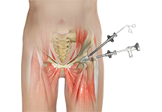
Hip Arthroscopy
Hip arthroscopy, also referred to as keyhole or minimally invasive surgery, is a procedure in which an arthroscope is inserted into your hip joint to check for any damage and repair it simultaneously.
Revision Hip Surgery
Revision hip surgery may be recommended when a previous hip arthroscopy or osteotomy has failed to resolve painful and debilitating hip symptoms.
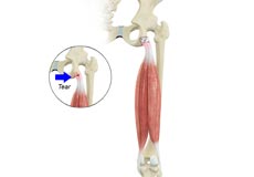
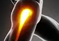
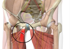
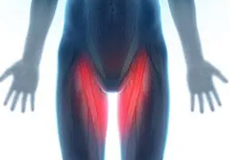
Proximal Hamstring Repair
These muscles help in extending your leg and bending your knee. Therefore, any damage to the hamstring muscle group affects both hip and knee movements.
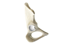
Acetabular Labrum Repair or Reconstruction
The acetabular labrum is a strong fibrocartilaginous tissue lining around the socket of the hip joint. It is an important structure that can cause significant pain and disability when it is damaged or torn. During arthroscopic surgery, the labrum is repaired or reconstructed with tendon tissue depending on the amount of damage that is seen.
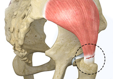
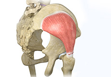
Gluteal Tendon Repair
Arthroscopic gluteus medius tendon repair is a minimally invasive surgical procedure employed for the treatment of a gluteus medius tendon tear, when the tear does not respond to conservative treatment.
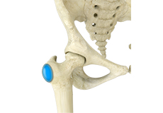
Core Decompression for Avascular Necrosis of the Hip
The hip joint is a ball and socket joint, where the head of the thighbone (femur) articulates with the cavity (acetabulum) of the pelvic bone.
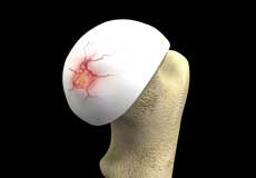
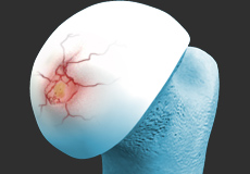
Hip Cartilage Repair
Hip cartilage is a white, tough, flexible tissue covering the ball (femoral head) and socket (acetabulum) of your hip joint. It acts as a cushion or shock-absorber and allows the bones to slide over one another by providing a smooth surface in the joint.
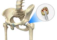
Hip Microfracture
Hip Microfracture is a marrow-stimulating technique that creates a network of small holes in the bone below the hip cartilage (subchondral bone). The goal of the procedure is to increase the blood supply to stimulate cartilage growth.
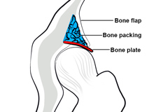
Hip Preservation Surgery
The hip is a ball and socket joint comprising of the femur (thigh bone) and the pelvic bone. The head of the femur (ball) articulates with a cavity (socket) called the acetabulum in the pelvic bone. To facilitate the smooth and frictionless movement of the hip joint, the articulating surfaces of the femur head and acetabulum are covered by spongy articular cartilage.
Physical Therapy for Hip
Physical therapy is an exercise program that helps you to improve movement, relieve pain, encourage blood flow for faster healing, and restore your physical function and fitness level. The main aim of physical therapy is to make your daily activities, such as walking, getting in and out of bed and climbing stairs, easier. It can be prescribed as an individual treatment program or combined with other treatments. Physical therapy is usually ordered to help you recover after certain surgeries, injuries and long-term health problems such as arthritis.
Core Muscle Repair
Core muscle injuries, also known as sports hernias or athletic pubalgia, can be effectively treated with an outpatient surgery called a core muscle repair.
Piriformis Tendon Release
Piriformis tendon release is an effective outpatient surgery when a tight piriformis tendon causes posterior hip or buttock pain that does not get better with physical therapy.
Lateral Femoral Cutaneous Nerve Decompression
Irritation of the lateral femoral cutaneous nerve, also known as meralgia paresthetica, can cause burning pain and numbness to the front of the thigh. Lateral femoral cutaneous nerve decompression is an effective outpatient surgery to relieve these symptoms.
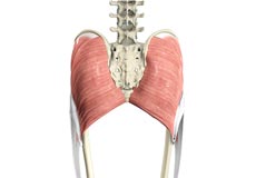
Gluteus Maximus Transfer
Gluteus maximus transfer, also referred to as gluteus maximus flap transfer (GMT), is a surgical technique to treat severe pain, limp, and instability associated with loss of the abductor portions of the gluteus medius and gluteus minimus muscles of the hip secondary to total hip arthroplasty or primary abductor disruption.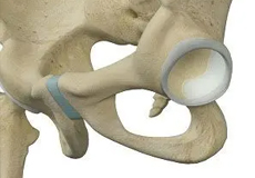
Hip Labral Augmentation
The hip joint is one of the body's largest weight-bearing joints and is the point where the thighbone (femur) and pelvis (acetabulum) join. It is a ball-and-socket joint in which the head of the femur forms the ball, and the pelvic acetabulum forms the socket.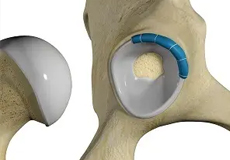
Hip Labral Reconstruction
Hip labral reconstruction is a surgery that involves the use of a graft to replace the damaged portion of a hip labrum.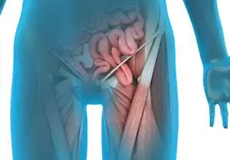
Sports Hernia Repair
Sports hernia repair is a surgical procedure performed to treat a sports hernia, also known as athletic pubalgia.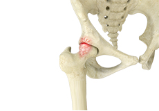
Femoroacetabular Impingement
Femoroacetabular impingement (FAI) is a condition characterized by excessive friction in the hip joint from the presence of bony irregularities. These cause pain and decreased range of hip motion.
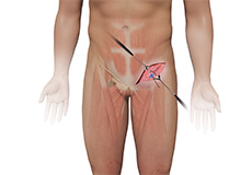
Athletic Pubalgia
Athletic pubalgia, also known as sports-hernia and Gilmore’s groin, is a condition characterized by chronic pain due to damage or strain to the soft tissues in the groin and pelvic region. This condition is mostly observed in athletes undergoing vigorous movements where there is twisting or a sudden change in direction. Although this condition is not a true hernia, it can lead to one.

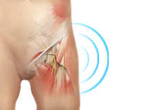
Deep Gluteal Pain Syndrome
Deep gluteal pain syndrome is deep posterior hip or buttock pain, with or without pain radiating down the thigh. This syndrome can be caused by several conditions including but not limited to an entrapped or irritated sciatic nerve, tight piriformis tendon, and chronic proximal hamstring tendinitis.
Meralgia Paresthetica
Meralgia paresthetica is a painful condition caused by entrapment or irritation of the lateral femoral cutaneous nerve (LFCN). The lateral cutaneous nerve provides sensation to the front of the thigh, and irritation of this nerve can cause burning pain and/or numbness along the front of the thigh.
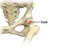
Femoral Cam Deformity
Femoral cam deformity, also referred to as cam impingement, is an irregularity or malformation of the ball at the top of the femur, or thigh bone.
Acetabular Pincer Deformity
Acetabular pincer deformity, also referred to as pincer impingement, is an abnormality of the acetabulum (hip socket) where the acetabulum is excessively deep or over covers the femoral head, resulting in impingement of the femoral neck and rupture of the labrum.
Failed Prior Hip Arthroscopy
This is the most common reason for a revision. Part of the labral repair surgery is to shave excessive bone around the hip that may be causing a labral tear. Sometimes, a surgeon may not remove enough bone, meaning that the cause of your labral tear was not adequately addressed.
Hip Dysplasia (Acetabular Dysplasia)
Developmental dysplasia of the hip (DDH) or hip dysplasia is a condition that is seen in infants and young children because of developmental problems in the hip joint. The femur (thighbone) partially or completely slips out of the hip socket leading to dislocation at the hip joint. It is most common in the first-born baby with a family history of the disorder.
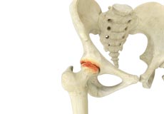
Hip Arthritis
Osteoarthritis, also called degenerative joint disease, is the most common form of arthritis. It occurs most often in the elderly. This disease affects the tissue covering the ends of bones in a joint called cartilage. In osteoarthritis, the cartilage becomes damaged and worn out, causing pain, swelling, stiffness and restricted movement in the affected joint. Although osteoarthritis may affect various joints including the hips, knees, hands, and spine, the hip joint is most commonly affected. Rarely, the disease may affect the shoulders, wrists, and feet.
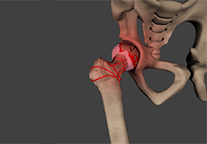
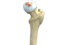
Femoral Head Avascular Necrosis
Hip osteonecrosis occurs due to disruption of the blood supply to the highest part of the thigh bone (femoral head). Due to lack of nourishment, the bone tissue of the femoral head dies and gradually collapses, which may further lead to degeneration of the underlying cartilage.
Hip Bursitis
Hip bursitis is a painful condition caused by the inflammation of a bursa in the hip. Bursae are fluid-filled sacs present in the joints between bone and soft tissue to reduce friction and provide cushioning during movement.
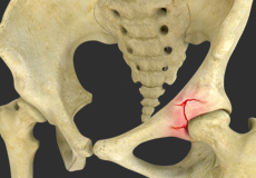
Labral Tear
The hip joint is a ball and socket joint in which the head of the femur is the ball and the acetabulum forms the socket. The labrum helps to deepen the socket and provide stability to the joint. It also acts as a cushion and enables smooth movement of the joint.
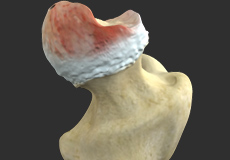
Torn Hip Cartilage
Hip cartilage is a white, tough, flexible tissue covering the ball (femoral head) and socket (acetabulum) of your hip joint. It acts as a cushion or shock-absorber and allows the bones to slide over one another by providing a smooth surface in the joint.
Legg-Calve-Perthes Disease
Legg-Calve-Perthes disease (LCPD) or Perthes disease is a disorder of the hip that affects children, usually between the ages of 4 and 10. It usually involves one hip, although it can occur on both sides in some children. It occurs more commonly in boys than girls.
Slipped Capital Femoral Epiphysis (SCFE)
Slipped capital femoral epiphysis (SCFE) is an unusual disorder of the hip where the ball at the upper end of the thighbone (femur) slips in a backward direction. This is caused due to weakness of the growth plate. This condition is commonly caused during accelerated growth periods such as the onset of puberty.
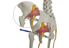
Piriformis Syndrome
Piriformis Syndrome is an uncommon, rare neuromuscular condition caused by the compression of the sciatic nerve by the piriformis muscle. The sciatic nerve is a thick and long nerve that passes below or through the piriformis muscle and goes down the back of the leg and finally ends in the feet in the form of smaller nerves.
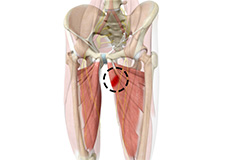
Core Muscle Injury: Chronic Hip Adductor/Groin Pain
Groin injuries are injuries sustained by athletes during sports activity. Groin injuries comprise about 2 to 5 percent of all sports injuries. The most common kind of groin injury is a groin strain or a pulled groin muscle. Typical groin injuries vary based on athlete’s gender, age, and sports, with most common groin injuries occurring in certain sports such as football with a prevalence rate as high as 12-16%.
Hip Muscle Strains, Tears, or Tendinitis
Tendons are strong connective tissue structures that connect muscle to bone. Hip tendonitis is a condition associated with degeneration of the hip tendons. This condition is mainly caused due to strain on the tendons which may occur due to overuse or biomechanical problems.
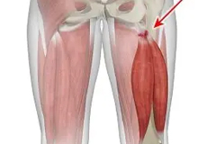
Hip Adductor Injuries
Hip adductors are the group of muscles on the inner side of your thigh that enable adduction or the ability to bring the thighs together.
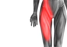
Hamstring Injuries
The hamstring is a group of three muscles that run along the back of the thigh from the hip to the knee. Hamstring injuries occur when these muscles are strained or pulled. They are common in dancers and athletes of all sorts including runners and those who play football, soccer, basketball, tennis, etc.
Hip Flexor Strain
A hip flexor strain is an overuse injury to the flexor muscles of your hip and can range from a minor stretch injury to a complete tear of the muscle fibers or tendons.
Hip Flexor Pain
Hip flexor pain is a distressing feeling or discomfort noted in the hip and/or groin region that can make everyday activities, such as going up and down the stairs or lifting your leg to tie a shoe extremely difficult and painful and can severely limit your activity and mobility.
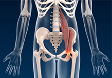
Iliopsoas Tendinitis
Iliopsoas Tendinitis also referred to as snapping hip syndrome, is an inflammation of the iliopsoas tendon or the surrounding area. The iliopsoas is the hip flexor tendon located over the front of the hip socket. The term snapping hip describes the sound made, a snap or click, that occurs with certain hip movements including flexion, extension, and rotation of the hip.
Hip Abductor Tear
Hip abductors are a major group of muscles found in the buttocks. It includes the gluteus maximus, gluteus medius, gluteus minimus, and tensor fascia lata muscles.
Gluteal Tendon Tear
The gluteal muscles (situated in the buttocks) are necessary for the stability and movement of the hip joints. The tendons of two gluteal muscles (gluteus medius and gluteal minimus) are attached at the outer hip region and are often called the “rotator cuff of the hip.” These tendons may be subject to injury or tearing due to various reasons. Since these gluteal muscles are involved in abduction (movement of your leg away from the midline of the body), the tears are also called abductor tendon tears.
Snapping Hip Syndrome
Snapping hip syndrome is a condition in which you hear or feel a snapping sound in the hip when you swing your legs, run, walk or get up from a chair. The sound can be experienced in the back, front or side of the hip.
Hip Dislocation
The hip joint is a “ball and socket” joint. The “ball” is the head of the femur or thighbone, and the “socket” is the cup-shaped acetabulum. The joint is surrounded by muscles, ligaments, and tendons that support and hold the bones of the joint in place. Hip dislocation occurs when the head of the femur moves out of the socket. The femoral head can dislocate either backward (posterior dislocation) or forward (anterior dislocation).
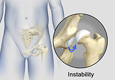
Hip Instability
Injury or damage to these structures can lead to a condition called hip instability when the joint becomes unstable.
Hip Ligament Injuries
Injuries to the hip ligaments are commonly called a hip sprain and can range from minor tears of the ligaments to more serious injuries involving the hip muscles, tendons or bone.
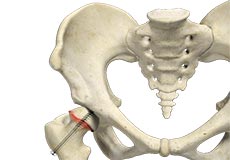
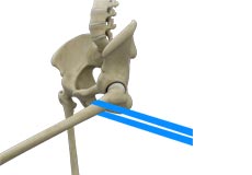
Stress Fractures of the Hip
Stress fractures of the hip are a break in the upper part of the thigh bone (femur) that fits into the socket of the hip joint. It can occur in any part of the hip, however, it mostly occurs just below the ball of the ball-and-socket hip joint called the femoral neck.
Transient Osteoporosis of the Hip
Transient osteoporosis of the hip is a rare condition that causes temporary bone loss in the upper region of the thighbone (femur). It occurs most often in young or middle-aged men of the age groups 30 to 60, and women in their later stages of pregnancy or early postpartum (following childbirth). It is characterized by abrupt onset of pain that increases with activity.
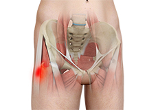
Gluteal Strain
A gluteal strain is a condition characterized by a partial or complete tear of the gluteus muscles, also known as the buttocks. The gluteus muscles are a group of strong muscles present at the back of the pelvis.
Groin Strain
A groin strain or groin pull is an overuse injury to the adductor muscles that run along the inner thigh and pubic region. They help draw the leg inwards and stabilize the torso. A groin strain may result in pain and loss of movement.
Groin Injuries in Athletes
Groin injuries are injuries sustained by athletes during sports activity. Groin injuries comprise about 2 to 5 percent of all sports injuries.
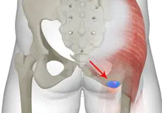
Ischial Bursitis
Bursae are fluid-filled sacs present between the tendons and bones to help facilitate smooth movement.
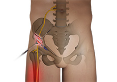
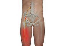
Sciatic Nerve Compression
The sciatic nerve is a large nerve that originates in the lumbar spine or lower back, running along the hip and back of the leg and branching off at the knee.
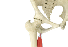
Partial Hamstring Tears
The hamstring is a group of three muscles known as semimembranosus, semitendinosus, and biceps femoris that run along the back of the thigh from the hip to the knee and help you extend your hip and bend your knee.
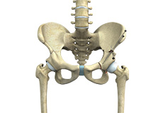
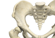
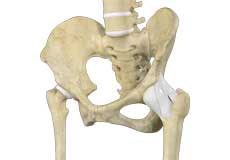
Hip Joint
The hip joint is the largest weight-bearing joint in the human body. It is also referred to as a ball and socket joint and is surrounded by muscles, ligaments, and tendons. The thigh bone or femur and the pelvis join to form the hip joint.
Any injury or disease of the hip will adversely affect the joint's range of motion and ability to bear weight.
The hip joint is made up of the following:
- Bones and joints
- Ligaments of the joint capsule
- Muscles and tendons
- Nerves and blood vessels that supply the bones and muscles of the hip
Bones and Joints
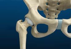
The hip joint is the junction where the hip joins the leg to the trunk of the body. It is comprised of two bones: the thigh bone or femur and the pelvis which is made up of three bones called ilium, ischium, and pubis. The ball of the hip joint is made by the femoral head while the socket is formed by the acetabulum. The Acetabulum is a deep, circular socket formed on the outer edge of the pelvis by the union of three bones: ilium, ischium, and pubis. The lower part of the ilium is attached by the pubis while the ischium is considerably behind the pubis. The stability of the hip is provided by the joint capsule or acetabulum and the muscles and ligaments which surround and support the hip joint.
The head of the femur rotates and glides within the acetabulum. A fibrocartilagenous lining called the labrum is attached to the acetabulum and further increases the depth of the socket.
The femur or thigh bone is one of the longest bones in the human body. The upper part of the thigh bone consists of the femoral head, femoral neck, and greater and lesser trochanters. The head of the femur joins the pelvis (acetabulum) to form the hip joint. Next, to the femoral neck, there are two protrusions known as greater and lesser trochanters which serve as sites of muscle attachment.
Articular cartilage is the thin, tough, flexible, and slippery surface lubricated by synovial fluid that covers the weight-bearing bones of the body. It enables smooth movements of the bones and reduces friction.
Ligaments
Ligaments are fibrous structures that connect bones to other bones. The hip joint is encircled with ligaments to provide stability to the hip by forming a dense and fibrous structure around the joint capsule. The ligaments adjoining the hip joint include:
- Iliofemoral ligament: This is a Y-shaped ligament that connects the pelvis to the femoral head at the front of the joint. It helps in limiting the over-extension of the hip.
- Pubofemoral ligament: This is a triangular shaped ligament that extends between the upper portion of the pubis and the iliofemoral ligament. It attaches the pubis to the femoral head.
- Ischiofemoral ligament: This is a group of strong fibers that arise from the ischium behind the acetabulum and merge with the fibers of the joint capsule.
- Ligamentum teres: This is a small ligament that extends from the tip of the femoral head to the acetabulum. Although it has no role in hip movement, it does have a small artery within that supplies blood to a part of the femoral head.
- Acetabular labrum: The labrum is a fibrous cartilage ring which lines the acetabular socket. It deepens the cavity, increasing the stability and strength of the hip joint.
Muscles and Tendons
A long tendon called the iliotibial band runs along the femur from the hip to the knee and serves as an attachment site for several hip muscles including the following:
- Gluteals: These are the muscles that form the buttocks. There are three muscles (gluteus minimus, gluteus maximus, and gluteus medius) that attach to the back of the pelvis and insert into the greater trochanter of the femur.
- Adductors: These muscles are located in the thigh which helps in adduction, the action of pulling the leg back towards the midline.
- Iliopsoas: This muscle is located in front of the hip joint and provides flexion. It is a deep muscle that originates from the lower back and pelvis and extends up to the inside surface of the upper part of the femur.
- Rectus femoris: This is the largest band of muscles located in front of the thigh. They also are hip flexors.
- Hamstring muscles: These begin at the bottom of the pelvis and run down the back of the thigh. Because they cross the back of the hip joint, they help in extension of the hip by pulling it backward.
Nerves and Arteries
Nerves of the hip transfer signals from the brain to the muscles to aid in hip movement. They also carry the sensory signals such as touch, pain, and temperature back to the brain.
The main nerves in the hip region include the femoral nerve in the front of the femur and the sciatic nerve at the back. The hip is also supplied by a smaller nerve known as the obturator nerve.
In addition to these nerves, there are blood vessels that supply blood to the lower limbs. The femoral artery, one of the largest arteries in the body, arises deep in the pelvis and can be felt in front of the upper thigh.
Hip Movements
All of the anatomical parts of the hip work together to enable various hip movements. Hip movements include flexion, extension, abduction, adduction, circumduction, and hip rotation.